DICOM Photometric Interpretation vs. Color Doppler in PACS
Same symptom on your system?
Tell us your model and what it is doing. A GEPROBE engineer replies with a diagnosis path, part options and pricing — usually within 6 hours, with no obligation.
Chat on WhatsApp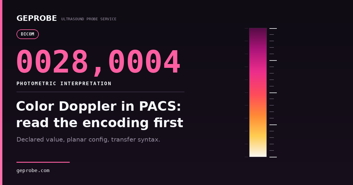
A color Doppler frame that reads orange in one viewer and blue-violet in the next sends a department straight to the probe. The faster answer is usually in its DICOM photometric interpretation.
That declaration, plus the three attributes that travel with it, explains most color complaints that never appear on the console itself.
This is a bench-side workflow, not a standards tour. First find the stage where color changes. Then split probe from console. Only then decide whether anyone should buy a part.
The order matters because the three questions have different owners, different clocks and very different price tags.
Locate the stage before you read a single tag
Most teams compare two things: what the sonographer saw and what PACS shows. That gap is too wide to act on.
Color Doppler that looks wrong in PACS but correct on the console is the classic encoding case. Color has to survive five stages, and the first stage that changes is the suspect.
| Stage | What you compare | If color changes here | Next move |
|---|---|---|---|
| 1. Live display | Probe image as scanned | Acquisition or console display path | Two-swap matrix below |
| 2. Console cine replay | Same saved loop, same frame | Console store or playback | Record software build, raise with console service |
| 3. Export re-imported | Your own export reopened on the same console | Exporter or transfer syntax | Request the exporter's conformance statement |
| 4. First receiving viewer | Same object in the receiving viewer | Decoder or display transform | Hand imaging IT the object identity and viewer build |
| 5. Second site viewer | Same object, different site | Fleet or version drift | Compare against that site's baseline object |
Table 1. One symptom, five stages, five different suspects.
With stages 1 and 2 clean and a change at stage 3 or 4, the question is encoding or decoding. No fault inside a transducer produces that pattern.
That single sentence saves more probes than any test we run.
Split probe from console in twenty minutes
Before touching DICOM at all, run two substitutions. Two swaps close most hardware questions and cost a fraction of a part.
- Same probe, different socket, color returns: connector pin path or channel, not the array.
- Different probe, same socket, fault follows the probe: probe-side, and worth a bench evaluation.
- Fault follows neither swap: console processing or display.
- Fault appears with both probes: something shared, often cable, connector seating or probe recognition.
One caution from our bench. A failing element shows as a band, a dropout line or a patch of empty color fill inside the vessel. It rarely repaints the whole hue map.
Whole-map hue changes point away from the array, which is exactly why a replacement probe so often arrives, tests clean and changes nothing.
If you want the threshold work behind that statement, our note on element dropout line tests walks through it.
What the standard fixes, and what it hands to the decoder
Photometric interpretation (0028,0004) declares how the stored samples should be read.
Samples per pixel (0028,0002), bits allocated (0028,0100), planar configuration (0028,0006) and the transfer syntax decide whether a decoder can realize it.
| Declared value | Samples / bits | What it implies |
|---|---|---|
| RGB | 3 / 8 | Planar value 0 or 1 sets byte order; naming RGB validates nothing |
| YBR_FULL | 3 / 8 | Luminance plus two chroma planes; US planar table fixes 1 for RLE |
| YBR_FULL_422 | 3 / 8 | Chroma subsampled; planar shall be 0; needs a JPEG codec |
| PALETTE COLOR | 1 / 8 or 16 | Indices plus three lookup tables, descriptors and data both |
| MONOCHROME2 | 1 / 8 | Grayscale; not by itself a lost-color fault |
Table 2. Read the declaration before suspecting hardware. Values per DICOM PS3.3 (2026d).
For ultrasound objects the module is strict about the pairing.
Samples greater than one must declare RGB for uncompressed or lossless syntaxes without defined color transforms, YBR_FULL_422 for JPEG lossy, and YBR_FULL or RGB for RLE (PS3.3 C.8.5.6.1.2 — reviewed 2026-11-08).
Three details catch people out. Planar configuration 0 interleaves red, green and blue per pixel; value 1 writes the red plane entire, then green, then blue (C.7.6.3.1.3 — reviewed 2026-11-08).
A palette descriptor whose entry count reads zero means 65,536 entries, not an empty table. And an RGB frame whose three samples are equal throughout is a legal gray image, so a colorless frame is not proof that color processing died.
There is also a real inconsistency in the text: the general pixel module and the ultrasound planar table do not fully agree on YBR_FULL order for uncompressed ultrasound objects.
Treat that as a question for the exporter's conformance statement. It is not a tag to edit.
Three comparisons that look like evidence and are not
Each of these produces a confident story and none of them survives a second look.
The live screen against a saved still compares two different objects. A screenshot against a DICOM file compares an appearance against samples.
Two frames of the same vessel compare two acquisitions with different gains and scale settings, so the hue difference may be nothing more than a change in the scale map.
Worse than all three is a habit: editing 0028,0004 so an old file opens. That rewrites the declaration, leaves nobody able to say what the exporter did, and turns a reportable software defect into a data incident.
Keep the original object, keep one unchanged copy, and work on a test copy.
Do not crate the probe until five things are true
This is the gate we ask referring departments to clear before a probe leaves their building. Each line is a yes or no, and a no stops the shipment.
- The color bar appears on a loop saved on the console, not only in PACS.
- Your own export, re-imported to the console, still shows color.
- A second probe on the same socket reproduces the fault, or clears the socket.
- The fault follows the probe onto a second system.
- Object identity, transfer syntax and viewer build are written down.
A probe that fails line two but passes line one is an export problem wearing a hardware costume. A lab will test it on a host system, find nothing, and return a no-fault report with freight attached.
Departments lose weeks to that loop, and the probe is never at fault.
The arithmetic that decides whether to buy a part
Cost arguments settle these disputes faster than any explanation of chroma subsampling. Here is the version we use with clinical engineering.
A = P + 2F + L*h + D*ds      (cost incurred if the part was not at fault)
T = L*ht + Xi                (cost of triage, incurred either way)
buy first only when q*A < T
P is the quoted part or probe price, F freight per leg, L your loaded labor rate, h internal handling hours, D clinical displacement per day, ds days the unit is out.
T pairs triage labor ht with viewer-side hours Xi. And q is your honest prior that the part is not the fault.
Run it with illustrative inputs, not our prices: P 6,000, F 200, L 60, h 8, D 900, ds 5.
That gives A = 6,000 + 400 + 480 + 4,500 = 11,380. Triage of three hours plus two viewer hours at the same rate gives T = 300.
Buy-first wins only when q × 11,380 < 300, so only when you are more than about 97 percent sure the probe is at fault. For a symptom that appears only in PACS, q sits near 1. The arithmetic never supports buying first.
Multi-site groups need one baseline object per site
Version drift is the quiet cause of "it only looks wrong at the north campus". One site's viewer converts chroma correctly, another sits a release behind.
Keep one anonymized color Doppler object in each site's test area. Record its hash, its declared attributes and the appearance your radiologists accept, then re-run it after any viewer, PACS, decoder or routing change.
The baseline never proves a file is conformant. It proves that a site's rendering moved, which is the question that actually arrives by email.
Where a viewer returns an empty study list, color is not the issue yet. Our note on DICOMDIR and viewer loading covers that earlier failure.
What we test, and what stays with your team
We repair probes and supply tested parts. Our evaluation mounts the probe on host ultrasound systems, so we can say whether the array, cable, connector or probe identification path is at fault.
We cannot sign off a viewer, and we will not edit a photometric tag to make a study open. Those belong to your imaging IT team and its software suppliers.
The intake is deliberately boring. A free evaluation with a diagnostic report comes first, followed by a flat-rate quote with repair-or-replace options.
Then OEM-standard restoration, functional test and imaging verification before return. Every probe ships with a test report and cosmetic checklist.
Current published terms, including the free 24-hour diagnostic, are on the repair service page; boards, cables, lenses and probe parts are in the parts catalog. Both were reviewed 2026-11-08.
Bring the object identity and the five-point gate with the probe. It shortens the conversation more than any description of the color.
Sources
- DICOM PS3.3 (2026d) C.8.5.6, US Image Module — reviewed 2026-11-08. Photometric, sample, bit-depth and planar tables for ultrasound.
- DICOM PS3.3 (2026d) C.7.6.3, Image Pixel Module — reviewed 2026-11-08. Defined photometric terms, planar order, palette lookup tables.
- GEPROBE repair services — reviewed 2026-11-08. Evaluation, quote and testing scope.
- GEPROBE ultrasound parts catalog — reviewed 2026-11-08. Part categories and availability basis.
All cost inputs in the worked example are illustrative assumptions, not GEPROBE prices. No statistics in this article were estimated; symptoms described are patterns we see in evaluation, not measured frequencies.
Related Articles
Ultrasound Transducer Useful Life: A Buyer's Decision Path
Cycle count, not manufacture date, decides what a used probe can still support. Screening fields, acceptance evidence, and arithmetic you can run.
Ultrasound USB DICOMDIR Viewer No Study: Four Layers
Files sit on the USB drive but the viewer lists no study. Work the four layers — media, naming, index, receiver — and know when the console is the fault.
Ultrasound DICOM Duplicate Instance Resend: Triage Order
A field order for an ultrasound DICOM duplicate instance resend: decide storage repeat, transport retry or viewer grouping before anyone sends it again.